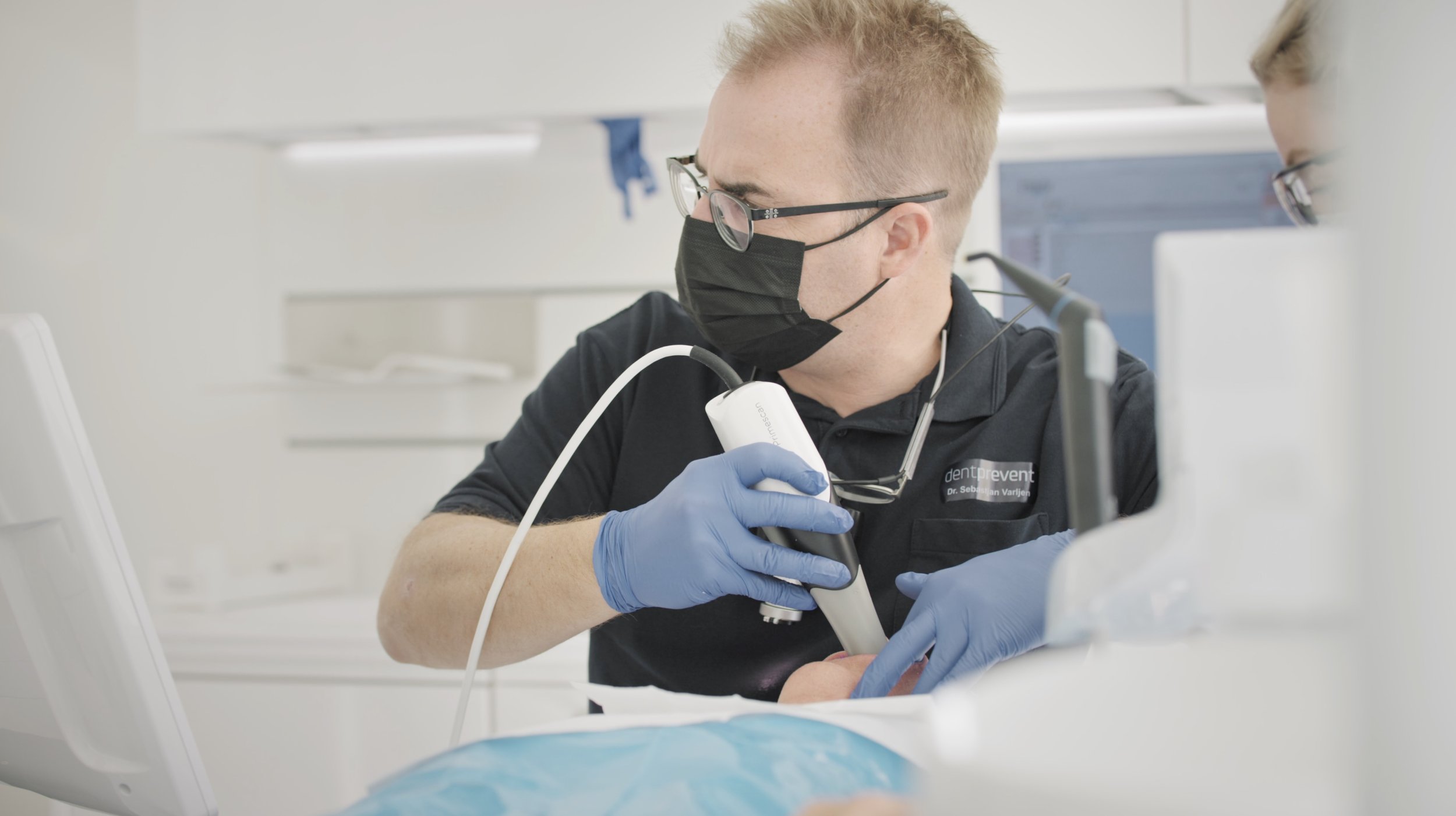
Why you should start intraoral scanning
Intraoral scanners help us dentists to capture detailed 3D images of our patients' dental features with incredible precision. The technology provides comprehensive details of teeth, gums, and occlusion. And patients are more than happy to avoid uncomfortable traditional impressions! Instead, they love to see the 3D image of their jaw appearing on the screen.
In our clinic, an intraoral scan is part of every patient examination. This practice ensures that we capture precise data for every treatment plan, enhancing the overall quality of care we provide.
By the end of this article, you will have a clear understanding of whether you should invest in a scanner (the short answer is: YES!), what to look for in a scanner, and how to integrate it into your daily workflow.
What is the difference between analog impressions and an intraoral scan?
Traditional dental impressions use materials like alginate or silicone to create physical molds of a patient's teeth. This often results in discomfort for the patients and potential inaccuracies and errors.
In contrast, intraoral scanners offer a faster, more precise, and comfortable experience. Dentists gently use a handheld wand with optical sensors to scan the patient’s mouth. By doing so, the scanner projects light onto the dental surfaces and captures the reflected images with high-resolution sensors, taking thousands of pictures per second. The accompanying software processes these images in real time, stitching them together to create a highly accurate 3D model of the patient's mouth, which can be viewed in real-time on a computer screen.
What are the benefits of intraoral scanners?
The transition from traditional impression methods to digital scanning marks a significant advancement in the dental world, as intraoral scanners provide numerous benefits for both practitioners and patients:
Intraoral scanners simplify the workflow for dental professionals, making the process easier and more efficient.
Digital impressions eliminate the need for physical impression materials, reducing costs and waste.
The scans provide highly accurate and detailed dental impressions to create precise dental restorations.
The scanning process is quick and non-invasive – Patients love the experience, which is much more pleasant than analog impressions.
The digital impressions can be sent to dental labs within seconds. Thanks to that, you will have faster turn-around times to get your dental appliances from your lab. The easy sharing process of the detailed data enhances the communication and collaboration with the lab.
Intraoral scanners capture detailed 3D images of teeth, gums, and occlusion. This precise data can improve the overall quality of care.
How do intraoral scans improve the treatment process?
Intraoral scans provide detailed, three-dimensional data of a patient’s teeth and surrounding tissues. They serve as an essential foundation for pre-treatment analysis.
At our clinic, we combine the intraoral scan with intra- and extraoral photos, a face scan, and jaw motion tracking analysis to create a comprehensive patient avatar. This thorough assessment allows us to set clear treatment goals and ensures precise and effective treatment planning.
Intraoral scans are essential to create accurate 3D models for various dental applications. You can immediately send these scans to your dental lab to produce the necessary dental work, such as 3D mock-ups, crowns, bridges and aligners. Alternatively, you can opt for in-house production by exporting the scan into CAD software like Exocad, designing your model, and printing it with your own 3D printer.
How to perform an intraoral scan:
A step-by-step guide
But how do you scan? Let’s walk through an intraoral scan.
Preparation
The first step is preparation. Make sure your scanner handpiece and mirror are clean and dry. A clear view is key to capturing all the details. We recommend to use a new single-use sleeve.
It's important to explain what you're doing to the patient. They need to feel comfortable and know what to expect. By making sure the mouth is clean and not too wet, you set the stage for a smooth scan.
Scanning process
For a good scan, start at one end of the upper jaw. Scan the palatal surface of the teeth and move smoothly over each tooth surface, the occlusal surfaces and the buccal area.
Include soft tissue and a full view of the palate: A fully scanned palate will help to align the model later in the lab software.
Repeat the process for the lower jaw. Try to capture the vestibulum so that the jaw is completely scanned. Finally, scan the bite from both sides. This step unites the jaws in static occlusion and gives us a snapshot of the patient's final bite position.
Scan review
Review the full scan on your computer screen: Cut away the structures you don't need, such as the cheeks and lips. Erasing these areas helps to reduce the file size and the 3D model is rendered in no time.
You can also easily rescan any areas to fill in missing data. But you shouldn't need to do this very often, as the lab software can fill in some minor gaps. After the final check, you are ready for approval: You can be confident that your digital impression is accurate and ready for the next steps.
Calculating the 3D model
The computer calculates the static 3D model, aligning it with the bite information in the final natural occlusion. If you want to change the occlusion, you can put something in between, like a deprogrammer or wax plate.
The computer creates the model in a new vertical dimension of occlusion (VDO). This shows us the relation between the jaws, also called the intermaxillary relation (IMR), which you can define or adapt according to your therapeutic needs. For example, in an abrasion case, if you want to perform a bite lift.
When you're happy with the final 3D scan of the upper and lower jaw, it's ready for export.
File management
Save the file in the appropriate format, typically an STL or PLY file. STL is monochromatic and has no color information, whereas PLY contains full color.
You can now upload the file to a dental cloud to create a digital patient avatar or share it with your external dental lab for 3D printing. It all depends on your workflow.

Want to learn more about digital dentistry?
Sign up for a free PDF guide
on digital dentistry!
Which intraoral scanner should I buy?
The technology of intraoral scanners has evolved significantly in recent years. There are a lot of compact, user-friendly and accurate options on the market. When choosing an intraoral scanner, consider key features such as accuracy and resolution, ease of use, ergonomics, software compatibility and integration, as well as costs.
Here is a list of scanners that we can recommend:
Planmeca Emerald S (approx. $10,000)
The Planmeca Emerald S scanner is fast and lightweight, making it easy to use. It captures vibrant and accurate digital impressions quickly and integrates smoothly with Planmeca’s comprehensive digital solutions.
Medit i500 (approx. $12,000)
The Medit i500 is known for its affordability and high resolution. It’s user-friendly, with a simple interface and great customer support. This makes the model a good fit for dental clinics that are new to digital scanning.
3Shape TRIOS 4 (approx. $20,000)
The TRIOS 4 by 3Shape provides superior accuracy and integrates seamlessly with a wide range of software solutions. It also provides advanced features such as caries detection and patient monitoring.
Dentsply Sirona Primescan (approx. $35,000)
The Primescan by Dentsply Sirona is renowned for its unparalleled accuracy and detail. It captures 50,000 images per second, creating a million 3D data points every second, ensuring extremely precise scans. The advanced software produces sharper details and higher accuracy compared to other models.
iTero Element 5D (approx. $48,000)
The iTero Element 5D scanner is a top-tier device offering comprehensive imaging capabilities, including 3D, intraoral color, and NIRI technology for detecting interproximal caries. It’s designed for high precision and efficiency in dental diagnostics and treatment planning.
A good intraoral scanner is the foundation for high-quality dental scans. While entry-level scanners like the Medit i500 may save money initially, precise scanning data is critical for every subsequent step in your treatment process.
Accurate scans ensure flawless fittings and superior restorations: This directly impacts treatment success and patient satisfaction.
For instance, while the Medit i500 excels in resolution on paper, Primescan’s advanced software produces sharper details with higher accuracy. With this level of detail, you can achieve the best possible treatment outcomes for your patients. This is why we decided to invest in a Primescan.
What do I have to consider if I want to start intraoral scanning?
High initial investment costs
The initial cost of an intraoral scanner can be quite high. So it is important to know that the investment is worthwhile. And it is! Integrating an intraoral scanner into your practice offers a remarkable return on investment and, more importantly, a significant return on time. For example, investing in a Dentsply Sirona scanner for €35,000 can quickly pay off: The scan can be performed by a dental assistant and typically takes about 15 minutes - or even less if it is done regularly. If you charge €100 per intraoral scan and perform 8 scans per week, you will break even in just 10 months.
The learning curve for staff
Buying a new device means that the staff will need to learn how to use it effectively. Many manufacturers offer resources like online tutorials to help you get started with their scanners. This ensures that your staff becomes proficient quickly and confidently.
If you are interested in a comprehensive training program on various digital tools and workflows in dentistry, you can attend our hands-on course. Among other things, you and your colleagues will receive hands-on demonstrations, scanning assistance and practical tips.
Compatibility of digital files with dental labs
If you are going to invest in an intra-oral scanner, don't overlook the fact that your dental lab should be able to receive and process the data. The digital files must be compatible with your lab. Use scanners that support open standard file formats, such as PLY or STL, to ensure broad compatibility. This way, you should be able to work easily and efficiently with any digital dental lab of your choice.
Quality control
Maintaining consistent, high-quality results with every scan is extremely important. To achieve this, implement strict quality control measures such as standardized scanning procedures, regular quality checks and best practices. An internal guide with a checklist for your team members helps to keep everyone on the same page. Don't forget to provide ongoing staff training, as well as regular calibration and maintenance of the scanner.
The upgrade to intraoral scanners:
A smart choice for your dental clinic
Investing in an intraoral scanner is a smart and strategic decision for any dental clinic aiming to improve patient care and streamline workflows. This technology is a great way to get started with digital dentistry and modernize your practice.
With a range of scanners on the market to suit different needs and budgets, every dental practice can find a suitable option to enjoy digital scanning.
Whether you start with an entry-level model or opt for a high-end scanner: Moving to digital impressions will undoubtedly improve your practice's efficiency and patient satisfaction. By integrating this technology into your daily operations, you can achieve better treatment outcomes and provide a more pleasant experience for your patients, positioning your practice at the forefront of dental innovation.


